Head & Neck Case Review
February, 2020
History
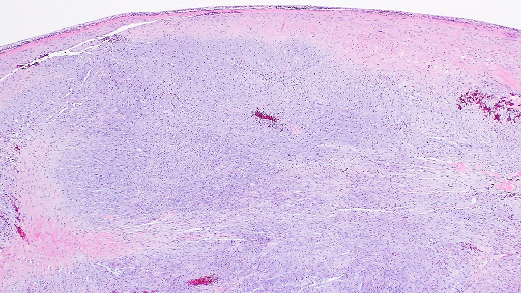
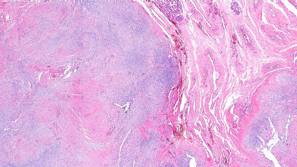
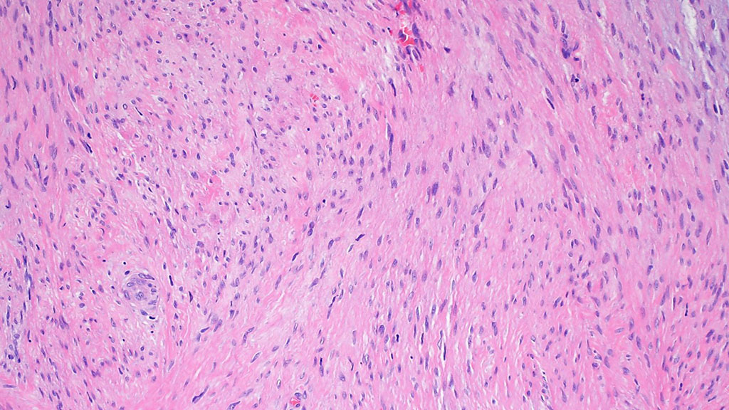
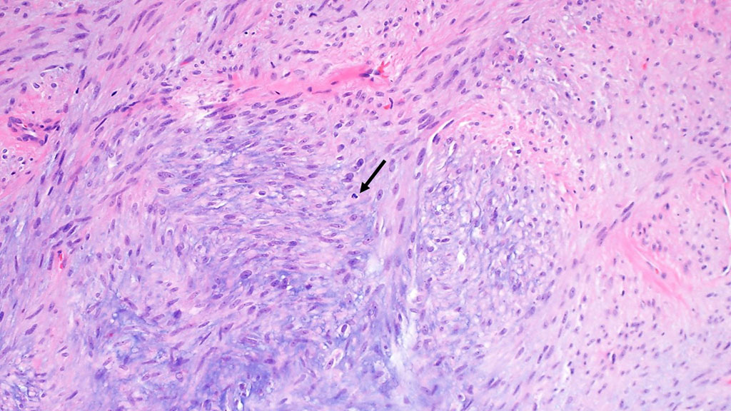
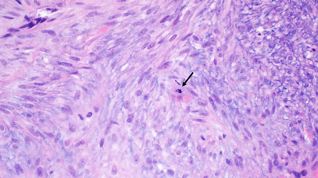
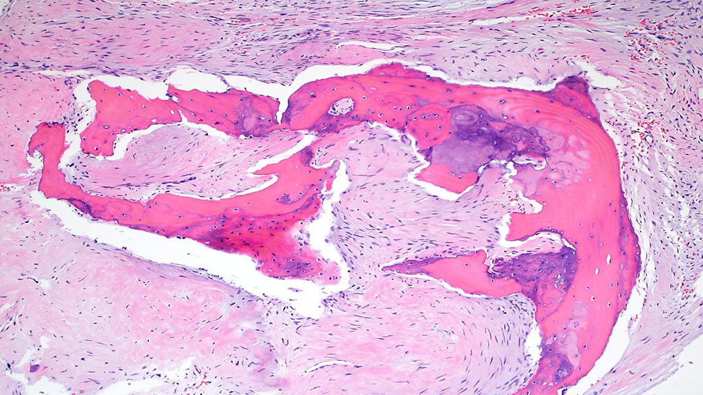
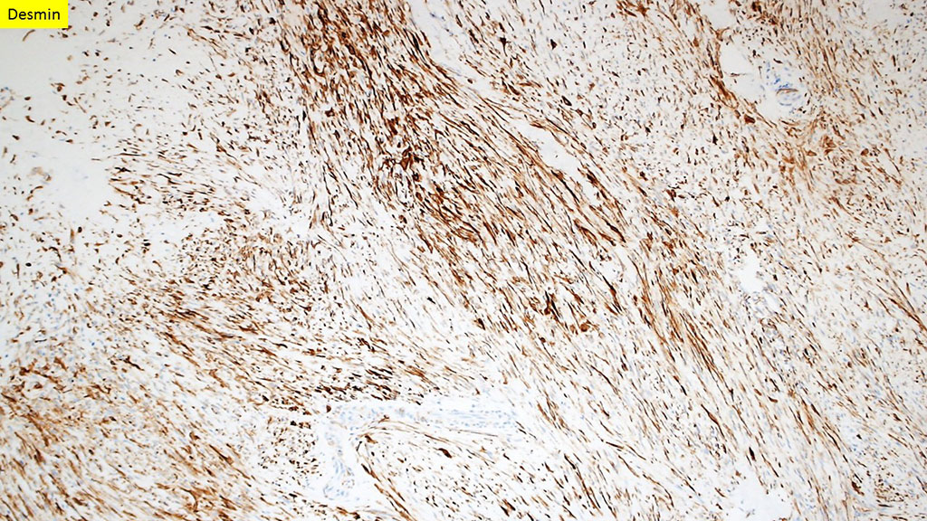
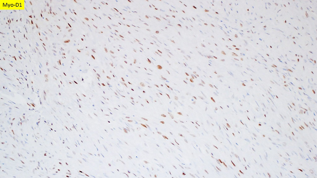
A 73-year-old man presented with right nasal obstruction and epiphora for about a year. On nasal endoscopy a smooth submucosal mass attached to the anterior part of the inferior turbinate and extending to the septum was identified. CT imaging also confirmed the presence of an ill-defined soft tissue mass confined to the nasal cavity. The patient subsequently underwent resection. H&E slides and immunostains are shown. The following stains were negative: cytokeratin AE1/AE3, CD34, S-100, SOX10, and myogenin.
Images
Quiz
Q1. The following genetic syndromes have been associated with this disease in children except:
- A. Li-Fraumeni syndrome
- B. Costello syndrome
- C. Neurofibromatosis type 1
- D. Beckwith-Wiedemann syndrome
- E. Cowden syndrome
Q2. The most common site of disease outside the Head and Neck is the paratesticular region.
- A. True
- B. False
Diagnosis
Spindle Cell Rhabdomyosarcoma.
Sinonasal rhabdomyosarcoma is a rare malignant mesenchymal tumour with skeletal muscle differentiation. The peak incidence is in the first decade of life, with no significant gender predilection. The most commonly involved sites in the Head and Neck (H&N) are the paranasal sinuses, followed by the nasal cavity. Outside the H&N, it is most often encountered in the paratesticular region of children. Embryonal rhabdomyosarcoma is the most common subtype in the sinonasal cavity whereas the spindle cell subtype is rarely seen in this site.
Most lesions present as polypoid, ill-defined masses with smooth surfaces, often involving adjacent structures. Histologically, spindle cell rhabdomyosarcoma consists of proliferation of spindle cells with elongated nuclei and pale indistinct cytoplasm in a fascicular pattern. Occasional obvious rhabdomyoblasts may be seen. The stroma may variably be myxoid, fibrous, and/or hyalinized.
By immunohistochemistry, the tumor cells are positive for desmin, Myo-D1, and myogenin. Spindle cell carcinoma and spindle cell malignant melanoma are among the differential diagnoses and should be excluded by negative keratin and melanoma markers, respectively.
Genetically, most alveolar rhabdomyosarcomas harbor PAX3-FOXO1 fusion and a small subset PAX7-FOXO1. Pediatric spindle cell rhabdomyosarcomas, however, show NCOA2 rearrangement. Rhabdomyosarcomas may be associated with genetic syndromes including Li-Fraumeni syndrome, Costello syndrome, Neurofibromatosis type 1, and Beckwith-Wiedemann syndrome.
Overall, the prognosis is poor in the sinonasal region and complete resection is difficult because of the location.
References
- Nascimento AF, Fletcher CD. Spindle cell rhabdomyosarcoma in adults. Am J Surg Pathol. 2005;29(8):1106–1113.
- Smith MH, Atherton D, Reith JD, Islam NM, Bhattacharyya I, Cohen DM. Rhabdomyosarcoma, Spindle Cell/Sclerosing Variant: A Clinical and Histopathological Examination of this Rare Variant with Three New Cases from the Oral Cavity. Head Neck Pathol. 2017;11(4):494–500. doi:10.1007/s12105-017-0818-x
- Carroll SJ, Nodit L. Spindle cell rhabdomyosarcoma: a brief diagnostic review and differential diagnosis. Arch Pathol Lab Med. 2013;137(8):1155–1158. doi:10.5858/arpa.2012-0465-RS
Quiz Answers
Q1 = E. Cowden syndrome
Q2 = A. True
Contributor
Mitra Mehrad, M.D.
Assistant Professor
Department of Pathology, Microbiology and Immunology
Vanderbilt University Medical Center, Nashville, TN